

 Computer analysis of slides containing lung cancer tissue samples increases the accuracy of tumor classification and patient prognoses, according to a new study conducted at Stanford University School Of Medicine in Stanford, California.
An essential element of subjectivity exists in pathologists’ traditional histopathological assessments, which uses a light microscope to assess thin cross-sections of biopsied tumor tissue on a slide to determine there tumor’s grade and severity. The more abnormal a tumor’s tissue appears in terms of cell size and shape, among other criteria, the higher the grade it is assigned. The tumor’s stage is also estimated based on whether and where the cancer has spread in the body.
Stanford researchers observed that this classification system doesn’t always work well for lung cancer, especially in lung cancer subtypes of squamous cell carcinoma and adenocarcinoma, which can be difficult to distinguish from each other when examining tissue culture slides. Moreover, they note that the stage and grade of a patient’s cancer doesn’t always correlate with prognosis, which can vary widely. Computers can make more consistently accurate lung cancer tissue assessments, they said, helping clinicians to decide on treatment methodology and how aggressively it should be applied.
The research team confirmed in the study that computer identification of key disease-related attributes leads to more accurate differentiation between these two types of lung cancer, and to better predictions of patient survival than the conventional approach used by pathologists.
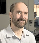 “Pathology as it is practiced now is very subjective,” said Michael Snyder, PhD, professor and director of the Stanford Center for Genomics and Personalized Medicine, in a press release. “Two highly skilled pathologists assessing the same slide will agree only about 60 percent of the time. This [computerized] approach replaces this subjectivity with sophisticated, quantitative measurements that we feel are likely to improve patient outcomes.”
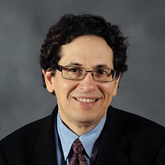 The research was published Aug. 16 in the journal Nature Communications, in a paper titled “Predicting non-small cell lung cancer prognosis by fully automated microscopic pathology,“ co-authored by Snyder, who shares the study’s senior authorship with Daniel Rubin,
  MD, an assistant professor of radiology and medicine. Graduate student Kun-Hsing Yu, MD, is the study’s lead author.
The investigators concluded that their results suggest that a machine-learning approach can predict prognosis of lung cancer patients, and contribute to precision oncology.
While this study’s focus was on lung cancer, the most prevalent cancer worldwide, the Stanford researchers believe their methods are relevant for other types of cancer.
“Ultimately this technique will give us insight into the molecular mechanisms of cancer by connecting important pathological features with outcome data,” Snyder said.
Drawing on 2,186 images obtained from patients with either adenocarcinoma or squamous cell carcinoma in the Cancer Genome Atlas national database, which also contained information on grade and stage assigned to each cancer and how long each patient lived after diagnosis, the researchers used the images to develop a computer software program able to recognize and identify nearly 10,000 individual traits — a much greater range of cancer-specific characteristics than can be detected by the human eye.
They focused particularly on a subset of cellular characteristics identified by the software that could most efficiently differentiate tumor cells from surrounding non-cancerous tissue, identify cancer subtype, and predict patient survival time after diagnosis. The team also validated the software’s ability to accurately distinguish short-term survivors from those who lived significantly longer, using another dataset of 294 lung cancer patients from the Stanford Tissue Microarray Database.
“We began the study without any preconceived ideas, and we let the software determine which characteristics are important,” said Snyder, who is the Stanford W. Ascherman Professor in Genetics. “In hindsight, everything makes sense. And the computers can assess even tiny differences across thousands of samples many times more accurately and rapidly than a human.”
The identification of previously unknown physical characteristics that can help to more accurately predict cancer severity is also likely to lead to a better understanding of molecular processes of cancer initiation and progression. For this reason, Snyder anticipates that this machine-learning system will be a useful tool in the emerging fields of cancer genomics, transcriptomics and proteomics — in which cancer researchers study DNA mutations, and gene and protein expression patterns, that lead to disease.
“We launched this study because we wanted to begin marrying imaging to our ‘omics’ studies to better understand cancer processes at a molecular level,” Snyder said. “This brings cancer pathology into the 21st century and has the potential to be an awesome thing for patients and their clinicians.”
The study was supported by the National Cancer Institute and the National Institutes of Health (grants U01CA142555 and 5U24CA160036-05), and also by Stanford’s Department of Genetics.
Sources:
Stanford University School of Medicine
Nature Communications