
 In a recent study entitled “Lineage-negative progenitors mobilize to regenerate lung epithelium after major injury” researchers identified a rare population of stem cells within the distal lung regions that are activated and migrate to major injury lung areas to mediate regeneration. The study was published in the journal Nature.
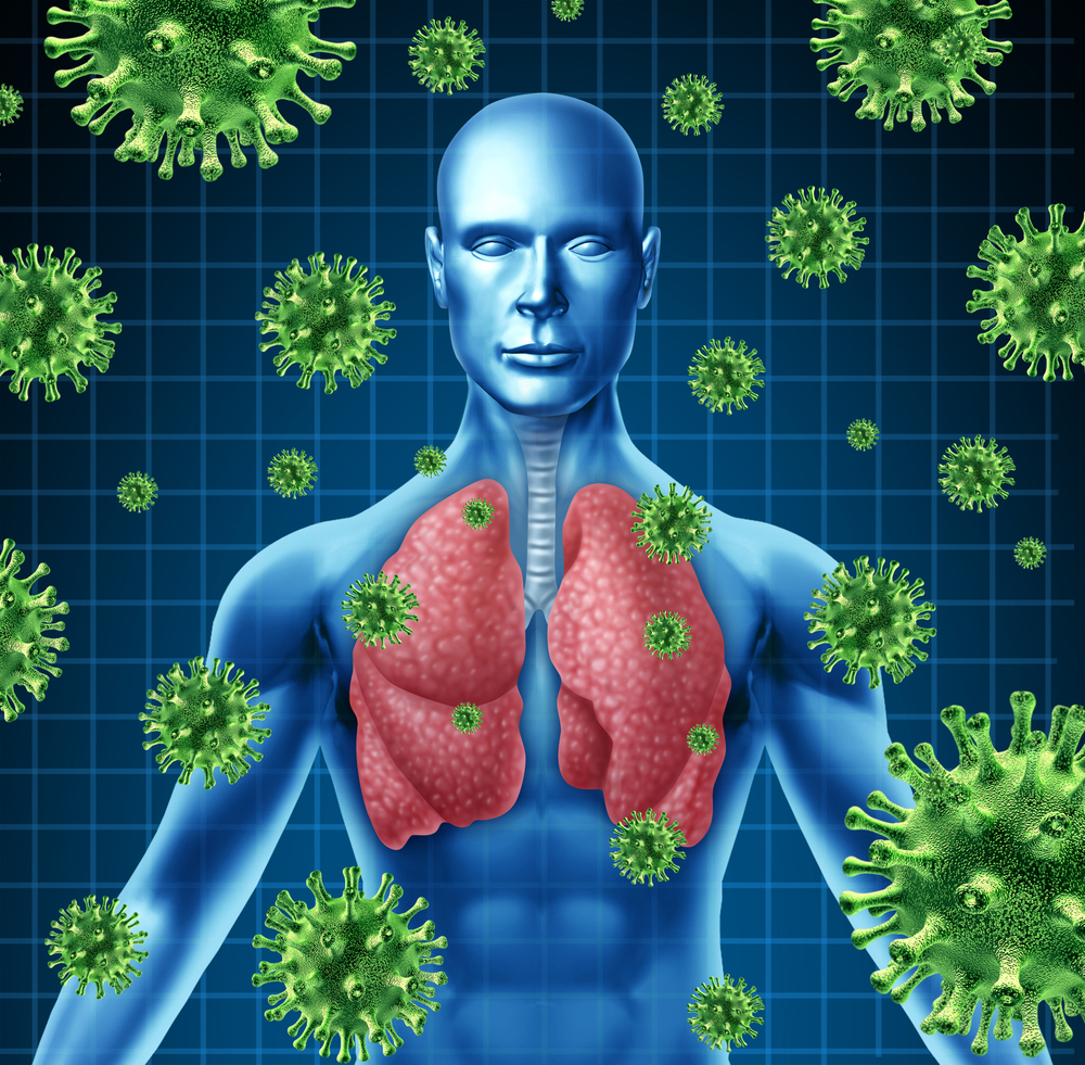
Influenza infection can cause severe loss of healthy lung tissue, however, it was observed that patients can recover from this massive loss of lung tissue. Notably, how this happens remains unclear. The lining of lungs (as well as, for example, the gastrointestinal tract and skin) is composed of epithelial cells. Upon injury, while skin and the gastrointestinal tract are quickly and constantly renewing their epithelia, this is not the case with the lungs. Current models for lung epithelial repair propose that when lung damage occurs, mature epithelial cells that survived the injury repair the epithelium.
In this study, a team of researchers at the University of California, San Francisco show that lung damage induced by influenza infection triggers the activation of a rare stem cell population within the lungs. These cells — termed, lineage-negative epithelial stem/progenitor cells (LNEPs) — are distinct from mature epithelial cells and once activated they proliferate and migrate to the site of injury. Here, injury-depleted mature cells areas are flooded with LNEPs cells that start to differentiate into mature epithelium.
[adrotate group=”7″]
The team performed more in depth-studies and discovered that the repair process was dependent on a Notch signaling pathway, specifically modulating the activation and differentiation of LNEPs cells. Notably, the authors found that after injury if Notch signals continues to persist, this in fact impaired the regenerative process mediated by LNEPs. In mice, the team observed that regions within the lungs with hyper-activated Notch signaling had honeycomb-like cysts, commonly seen in chronic lung infection patients, such as advanced idiopathic pulmonary fibrosis (IPF) and scleroderma. Accordingly, when the team analyzed human lung tissue from these patients, they observed that indeed lung cysts when present expressed high levels of Notch signaling.
The teams findings show that stem cells can repopulate damaged lung tissue, depending on the extent of the injury and regeneration and fibrosis is a delicate balance dependent on the action of Notch signaling. Another study focusing on lung epithelial regeneration was also published in the same issue of Nature and covered on Lung Disease News.