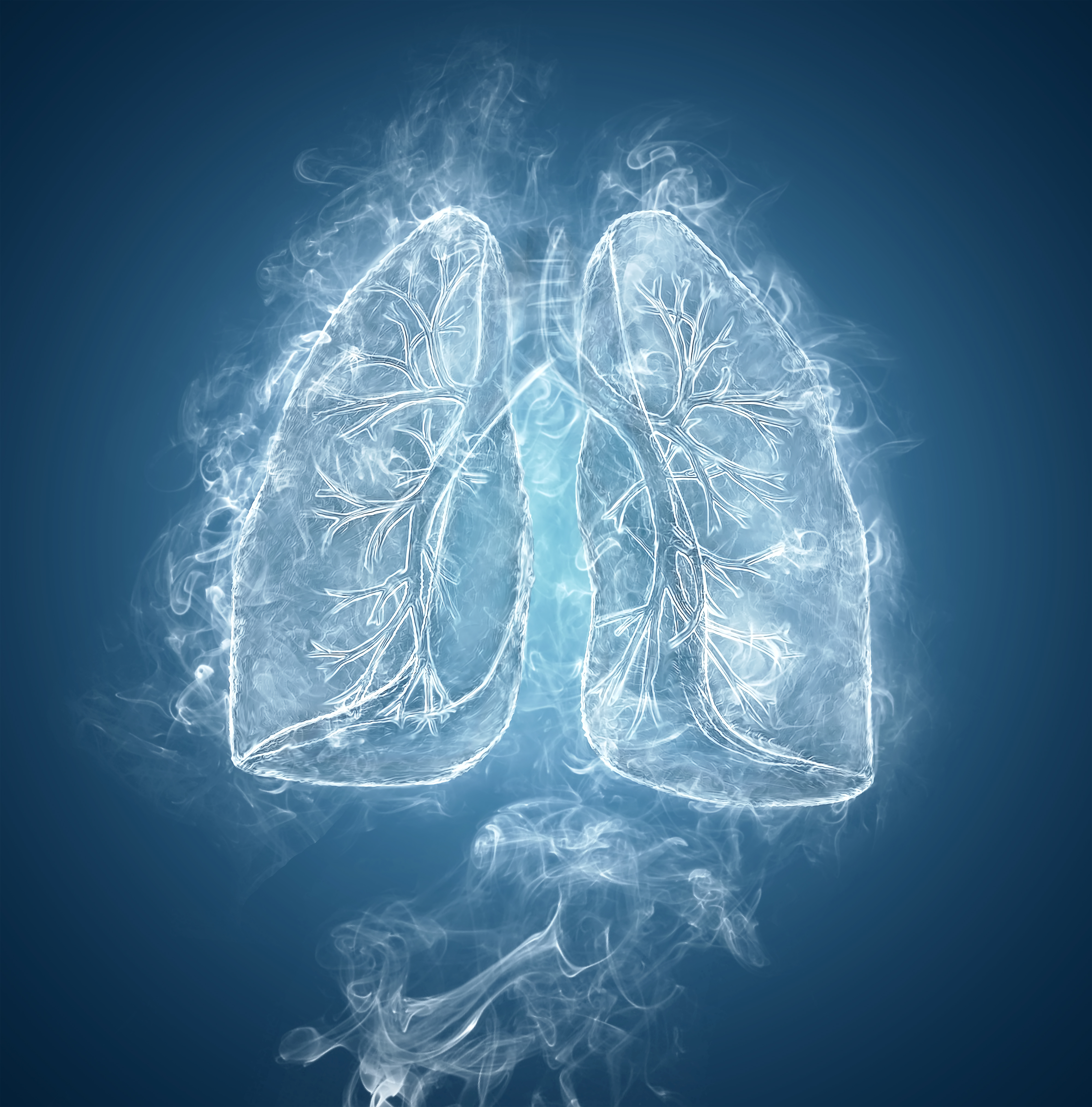
A multidisciplinary team of researchers from Arizona State University’s (ASU) Biodesign Institute, have recently released results from a study that may have important implications for the future of the science of lung tissue bioengineering. The study, entitled, “Recellularization of Decellularized Lung Scaffolds Is Enhanced by Dynamic Suspension Culture,” utilized a novel bioengineering strategy to address the problems associated with organ transplantation, such as, donor organ shortage and possible adverse immune response of recipients.
The study was conducted in the laboratory of Cheryl A. Nickerson, Ph.D. Professor of Life Sciences, School of Life Sciences, Center for Infectious Diseases and Vaccinology, The Biodesign Institute, ASU, and was published in the latest online edition of PLOS one. The aim of her laboratory’s research is to understand the effects of biomechanical forces on living cells (microbial and human), how this response is related to normal cellular functioning or infectious disease, and its translation to clinical applications. Dr. Nickerson and her team have developed several innovative model systems to study these processes including 3D organotypic models of human tissues that mimic the structure and function of cellular tissues and their application to study the host-pathogen interaction that leads to infectious disease.
Building designer lungs
Dr. Nickerson, and her team used an experimental mouse model to test a method in which a tissue scaffold is “reseeded” with stem cells that adhere to the engineered scaffold, grow and mature into the different cell types found in the functioning lung by various experimental cellular growth methods. The results of the study demonstrated a number of critical advantages to conducting the cell re-population process in a dynamic suspension culture (cells in liquid media), in which the cells are allowed to gently tumble as they re-seed the engineered lung scaffold.
In a university press release about the importance of the study, Dr. Nickerson, stated, “There is an urgent need for the development of lab-engineered lungs from patient stem cells that are suitable for both transplantation and as predictive models for biomedical research to probe the links between cell function and respiratory disease. This was an extraordinarily challenging but rewarding study that took years to complete, and we are excited that it will contribute to the current and growing body of knowledge in the field with potential downstream implications for regenerative medicine as well as identifying the underlying factors that may contribute to the transition of normal to fibrotic lung disease.”
This sentiment was shared by her colleague and co-author, Dr. Aurélie Crabbé, PhD, “Our study demonstrates that reseeding lung scaffolds under dynamic conditions could be beneficial for ex vivo lung engineering. Dynamic culture conditions enhanced cell growth and viability, and stimulated stem cells to differentiate into matrix-producing cells as compared to static conditions — both findings might help rebuilding lungs from cellular scaffolds.”
Dr. Crabbé, continued, “Tremendous progress has recently been made on the use of cadaveric lungs as building blocks for the generation of new lungs, and we are hopeful that continued advancements in this field will one day provide a new source of much-needed organs.”
When discussing the future implications of the study’s findings, Dr. Nickerson, stated, “Imagine, if we can one day create a fully functional normal or fibrotic lung in the lab, understand the reasons why it behaves the way that it does, learn the tipping points that cause it to transition either way, and test how preventative therapies work. The potential findings could have tremendous implications for diagnosis, treatment and prevention for a variety of respiratory disorders, including those due to infectious disease, cancer and environmental toxins.”

