
Scientists at the Helmholtz Zentrum München (HMGU) in Munich, Germany working in cooperation with the Ludwig-Maximilians-Universität Hospital and the Technische Universität München (TUM) report that they have for the first time tested X-ray dark-field radiography on a living organism to diagnose lung disease. This pioneering methodology enables production of highly detailed images of the lung. Conventional radiographic imaging is based on absorption of X-rays as they pass through the tissue. The newly developed X-ray dark-field radiography technique uses new technology to monitor wave changes during tissue transmission to create higher resolution images.
The team reports in the Investigative Radiology journal that this method shows promise in detecting diseases such as pulmonary emphysema at an earlier stage than it is currently available with conventional radiographic procedures generate images based on the absorption of X-rays as they pass through the tissue. The newly developed technique of X-ray dark-field radiography uses new technology to monitor wave changes during tissue transmission to create higher resolution images.
Detailed Images
With the aid of this new technique, the team from the HMGU, University Hospital Munich and TUM around Dr. Ali Önder Yildirim and Prof. Oliver Eickelberg of the Comprehensive Pneumology Center (CPC) , which is one of the centers of the German Center for Lung Research (DZL) , have achieved detailed images of soft tissue.
The CPC is a joint research project of the Helmholtz Zentrum München, the Ludwig-Maximilians-Universitt Clinic Complex and the Asklepios Fachkliniken Mnchen-Gauting, with the objective of conducting research on chronic lung diseases in order to develop new diagnosis and therapy strategies. The CPC maintains a focus on experimental pneumology with the investigation of cellular, molecular, and immunological mechanisms involved in lung diseases. The CPC is a site of the Deutsches Zentrum for Lungenforschung (DZL).
The CPC studies the immunopathology of COPD, specifically:
• Which subtypes of T-cell plays a role by the development of COPD pathogenesis?
• What is the specific function subset of CD4+T helper cell (e.g. Th17) and B cell in parenchymal lung tissue damage and small airway remodeling in response to cigarette smoke?
• What is the function of Protein Arginine Methyltransferases (PRMTs) in the pathogenesis of COPD?
The German Center for Lung Research (DZL) pools German expertise in the field of pulmonology research and clinical pulmonology. The association’s head office is in Giessen. The aim of the DZL is to find answers to open questions in research into lung diseases by adopting an innovative, integrated approach and thus to make a sizable contribution to improving the prevention, diagnosis and individualized treatment of lung disease and to ensure optimum patient care.
The Eickelberg laboratory investigates lung tissue remodeling using molecular and cellular mechanisms, leading to pathological remodeling of lung structure. The researchers note that normal remodeling is key to various chronic lung diseases, e.g. lung fibrosis, chronic obstructive lung disease (COPD), or bronchiolitis obliterans. State of the art genomic, biochemical, and cell biological methods, as well as mouse models, are used to elucidate remodeling processes contributing to such diseases. In particular, the Eickelberg lab is interested in signaling processes influencing epithelial cell plasticity, epithelial-mesenchymal crosstalk and extracellular matrix dynamics. The lab also explores biomarkers and molecular signatures of cells involved in disease onset or progression, and correlates these findings to human samples.
The study “Improved Diagnosis of Pulmonary Emphysema using in vivo Dark-Field Radiography” (M. Bech, A. Tapfer, A. Velroyen, A. Yaroshenko, B. Pauwels, J. Hostens, P. Bruyndonckx, A. Sasov, F. Pfeiffer. In-vivo dark-field and phase-contrast x-ray imaging. Scientific Reports, 2013; 3 DOI: 10.1038/srep03209) was conducted in cooperation with the Cluster of Excellence Munich-Centre for Advanced Photonics (MAP) .
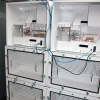
 The scientists used a small-animal scanner developed by Prof. Franz Pfeifer at the TUM to test X-ray dark-field radiography on a living organism. For their investigations, they evaluated and compared images of the lung. ”With X-ray dark-field Iradiography, structural changes in the lung tissue are visible at an early stage,” says the CPC/HMGU’s Dr. Yildirim.
At the MAP, physicists, chemists, biologists and medical experts are working on development of groundbreaking light-and laser-based particle sources. Since its foundation in 2006, the Cluster has been one of the leaders in laser science. All parameters of light – from its rate of propagation to its spectral composition – can now be precisely controlled. Scientists at MAP are developing and using new light sources to obtain detailed insights into the microcosmos by steering and tracing electrons with controlled light forces. In combination with innovative imaging techniques, new laser-driven secondary sources promise to improve the diagnosis and therapy of life-threatening diseases. Novel laser-powered particle sources also have the potential to greatly enhance the success rates of treatments for many types of cancer.
Early Detection Of Lung Disease
“Early detection of changes in the lung tissue will improve the diagnosis of lung diseases,” explains Dr. Felix Meinel from the Institute of Clinical Radiology at the University Hospital Munich. The clinical application, in particular the diagnosis of lung diseases such as pulmonary emphysema or pulmonary fibrosis, will now be tested in further studies.
Lung diseases are among the leading causes of death worldwide, and factors including genetics, lifestyle and environmental factors all play a role in their development. The work of the Helmholtz Zentrum München, the German Research Center for Environmental Health, focuses on the major common diseases with the aim of developing new approaches to their diagnosis, treatment and prevention.
Helmholtz Zentrum München investigates important common diseases which develop from the interaction of lifestyle, environmental factors and personal genetic background, focusing particularly on diabetes mellitus and chronic lung diseases.
Helmholtz Zentrum München is a research institution of the Federal Republic of Germany and the Free State of Bavaria. It is a member of the Helmholtz Association of German Research Centers. As the German Research Center for Environmental Health, Helmholtz Zentrum München Helmholtz Zentrum München investigates important common diseases which develop from the interaction of lifestyle, environmental factors and personal genetic background, focusing particularly on diabetes mellitus and chronic lung diseases, and pursues the goal of developing personalized medical approaches for the prevention and therapy of major common diseases such as diabetes mellitus and lung diseases. To achieve this, HZM investigates the interaction of genetics, environmental factors and lifestyle.
The Helmholtz Zentrum München has about 2,200 staff members and is headquartered in Neuherberg in the north of Munich, and is a member of the Helmholtz Association, a community of 18 scientific-technical and medical-biological research centers with a total of about 34,000 staff members.
Source:
Helmholtz Zentrum München (HMGU)
Image Credits:
HMGU
Comprehensive Pneumology Center (CPC)