
 Gregory Paulin and colleagues from Robarts Research Institute and The University of Western Ontario, Canada, presented results in an abstract of a pilot study aimed at examining the relationship between 3He MRI ventilation defects and well-established measurements of bronchiectasis in 15 patients aged 18-65 with Cystic Fibrosis or Non-CF Bronchiectasis.
Bronchiectasis is a chronic pulmonary disease characterized by damaged airways that cause abnormal dilation of the bronchi, leading to poor clearance and pooling of mucous in affected areas. Airway wall abnormalities result from chronic inflammation due to an inability to clear mucous secretions, which is common in both cystic fibrosis (CF) and non-CF subjects.
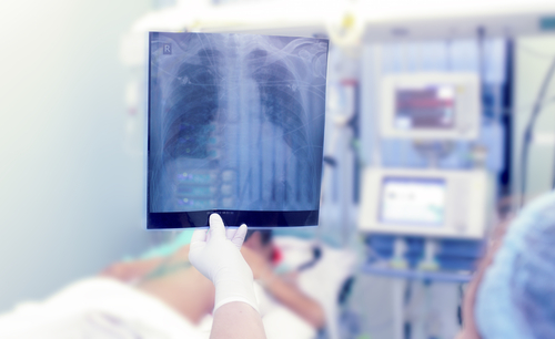
Evidence shows that hyperpolarized gas magnetic resonance imaging (MRI) provides an in-vivo assessment of regional gas distribution in the lung. Previous studies have shown that MRI has the advantage of showing exactly where regional functional abnormalities occur, and that heterogeneously distributed lung function abnormalities are associated with pulmonary function and symptoms.
The pilot study included 15 patients (non-CF and CF) with a diagnosis of bronchiectasis and a FEV1>25% that within 10 minutes of completion of post-salbutamol pulmonary function tests were evaluated with the 3He MRI, and after 30 minutes with CT chest. 3He MRI was analyzed for ventilation defect percent (VDP), a surrogate measurement of the normalized volume of the lung that is not ventilated. Thoracic CT volumes were also evaluated for the generation of quantitative airway measurements including airway wall area percent and lumen area, peribronchial thickening, and mucous plugs.
[adrotate group=”6″]
The results showed that pulmonary functional MRI with inhaled 3He provided visualization of regional ventilation and ventilation defects in patients with bronchiectasis. Using linear regression, the study found a relationship for FEV1 and 3He VDP. However, to enable potential treatment effects it is necessary to further test the spatial relationship between ventilation abnormalities with peribronchial thickening and mucous plugs.
This pilot study was funded by the Canadian Institutes of Health Research and is part of an investigation aiming to validate MRI measurements of airway function as intermediate endpoints for bronchiectasis therapy studies.