 Patients with bronchiectasis are intimately familiar with the disease’s vicious cycle of symptoms. Recurrent inflammation and infection, two hallmarks of the disease, lead to compromised mucociliary clearance, disallowing mucous removal and allowing further inflammation and infection in the bronchi. Despite these uncomfortable and hazardous symptoms, bronchiectasis is too often under diagnosed by clinicians, as cited by several sources, including the University of Chicago Medicine.
According to a recent study commissioned, under-diagnosis may be due largely to its grouping under chronic obstructive pulmonary disease (COPD). “The clinical features of the COPDs frequently overlap,” wrote Jane M. Braverman, PhD, in the article “Airway Clearance Indications in Bronchiectasis: An Overview.”
Contributing further to underdiagnosis is the fact that bronchiectasis is a manifestation of other airway diseases such as cystic fibrosis, meaning that bronchiectasis may simply be overlooked by a diagnosis of severe symptoms of cystic fibrosis. Other triggers of bronchiectasis include exposures to caustic fumes in the environment, lung infections, or blocked airways.
It may be surprising that bronchiectasis is underdiagnosed, as it was considered a common condition before the advent of antibiotics and immunizations against childhood diseases. After the mid 1950s, when immunization campaigns and antimicrobial agents became mainstream, childhood infection declined, as did the prevalence of bronchiectasis diagnoses. For a comparison, in 1940 a report indicated that 92% of patients with bronchiectasis died as a result of the disease, while in 1969 the percentage declined to 50%, and in the 1970s it was less than 10%.
[adrotate group=”6″]
Nowadays, approximately 110,000 people each year in the United States are treated for bronchiectasis, translating into a medical expenditure of $630 million a year. These treatments can only treat the symptoms of bronchiectasis, as there is no cure for the condition. The goal of treatment is to clear mucus from the lungs to end the vicious cycle and provide relief to patients. Yet in order to receive treatment, patients must first be diagnosed.
Researchers are aware that bronchiectasis is underdiagnosed, and they are identifying ways in which the disease can be brought to the front of clinicians’ minds when they are presented with the symptoms of bronchiectasis. At The University of Texas Health Center at Tyler Hospital and Clinics, a team of doctors conducted a retrospective study of 123 patients with well-documented bronchiectasis records to establish a “characteristic clinical picture of bronchiectasis… [that] should allow ready recognition of the disease.” The study, “Clinical, Pathophysiologic, and Microbiologic Characterization of Bronchiectasis in an Aging Cohort,” was published in the journal Chest.
Over half of the confirmed cases of bronchiectasis were identified using a CT scan, while a large percentage was diagnosed by bronchography or surgery. Looking at patient histories revealed that 70% of patients had an event that may have caused their bronchiectasis, such as pneumonia. Seventy percent also had chest crackles upon examination, while 34% had wheezing.
Delving into the more technical side of testing, pulmonary function tests identified airway obstruction in more than half (54%) of the patients. Samples of sputum generally tested positive for the pathologic microbial flora Pseudomonas aeruginosa. It may be valuable to examine the sputum of all patients with symptoms of bronchiectasis, as it may indicate superior treatments options, just as it does in the case of COPD — bearing in mind that the two diseases are not the same. By creating a clinical picture of bronchiectasis, doctors are more aware of the signs of disease, and this may help reduce the high rate of underdiagnosis.
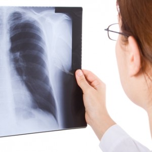
 The team at UTHC believes a reason for underdiagnosis resides “in the fact that few textbooks in pulmonary medicine portray bronchiectasis as a significant and/or common lung problem.” One book that is useful to describing bronchiectasis is The Little Black Book of Pulmonary Medicine, a compilation of diagnostic techniques and therapeutics for a wide range of pulmonary diseases, which are also described in detail. The book indicates that bronchiectasis can be diagnosed through high-resolution CT scanning of the chest or plain chest X-rays. Nealy all (91%) of the patients in the UTHC study showed fibrotic stranding and infiltrates in chest radiographs, which contributed to diagnosis. Prolonging diagnosis is hazardous to the patient, as chronic infection may lead to malnourishment due to high metabolic requirements.
“Many patients, if caught early enough and treated aggressively, can lead a fairly normal life,” indicated a representative from the University of Chicago Medicine’s Center for Advanced Lung Diseases. “Living with this disease process requires diligence and commitment.” Ultimately, the disease also requires a diagnosis. As researchers work to attain a greater understanding of the disease onset and symptoms, bronchiectasis may soon be higher on the list of possible diseases that cause patient symptoms, rather than considered a differential diagnosis.

