

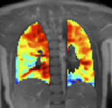
A research team from the Lerner Research Institute at the Cleveland Clinic recently discovered that a particular enzyme that is part of protein modification through a glucose metabolism pathway (HBP) might stimulate the proliferation of cells in the lung tissue of patients with idiopathic pulmonary arterial hypertension (IPAH).
The hexosamine biosynthetic pathway (HBP) operates as a sensor for metabolic flux and is a precursor for the glycosylation pathway. When activated, the HBP produces a sugar nucleotide called UDP-N-acetyl-glucosamine (UDP-GlcNAc), a substrate for hyaluronan (HA) and CMP-sialic acid synthesis, and for the O-linked ȕ-N-acetylglucosamine (O-GlcNAc) modification of proteins.
O-GlcNAc modification is a cellular process similar to protein phosphorylation with occupation of the same serine/threonine residues when the phosphate is removed. Usually, O-GlcNAc modification has an inverse relationship with phosphorylation and is central to the regulation of the target protein’s function.
In their study titled “O-GlcNAc Transferase Directs Cell Proliferation in Idiopathic Pulmonary Arterial Hypertension” recently published in the Journal Circulation, Jarrod Barnes and colleagues aimed to determine the relationship between glucose metabolism and smooth muscle cell proliferation in patients with IPAH.
They found that the hexosamine biosynthetic pathway (HBP) was unregulated in lung tissue from eight patients with IPAH compared with lung tissue from eight controls without the disease.
[adrotate group=”4″]
Specifically, the researchers found that activation of the HBP directly increased OGT levels and activity, triggering changes in glycosylation and PASMC proliferation. Partial knockdown of OGT in IPAH PASMCs resulted in reduced global O-GlcNAc levels and abrogated PASMC proliferation. The increased proliferation observed in IPAH PASMCs was directly impacted by proteolytic activation of the cell cycle regulator, host cell factor-1 (HCF-1).
The results indicate that proliferating cells in patients with IPAH are mainly using aerobic glycolysis to metabolize oxygen, because this pathway offers not only the energy but also the “building blocks” for quick proliferation.
The researchers examined OGT levels in 86 IPAH patients’ red blood cells (RBC), finding them to be significantly increased, as was O-linked GlcNAc protein modification. The results revealed that RBC OGT levels were associated with clinical severity, and were higher in patients with worse right heart stroke volume and 6-minute walk distance and with more severe functional status (New York Heart Association functional class). In the study, patients that had higher levels of OGT were 3.71-fold more likely to be hospitalized, to need lung transplants or to die during a median time of 24.5 months.
Based on the results, the research suggests an IPAH model considering the up regulation of HBP in proliferating cells that leads OGT to use UDP-GlcNAc to cleave HCF-1, causing an increase in PASMC proliferation.
“This model establishes a regulatory role for OGT in IPAH, sheds a new light on our understanding of the disease pathobiology, and provides opportunities to design novel therapeutic strategies for IPAH,” the researchers conclude in Circulation.